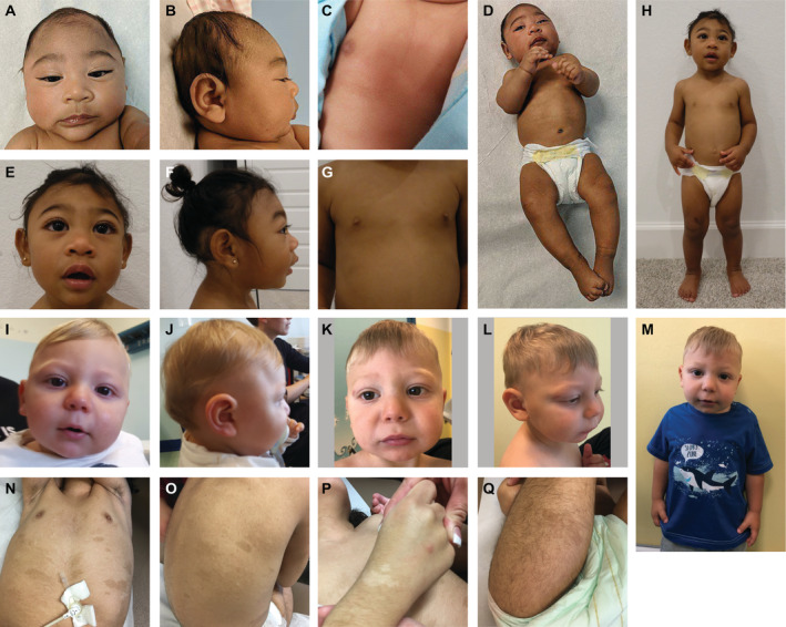
Figure 4.

Clinical photographs of individuals with ANKLE2 variants. Proband 9 at 4 months (A–D) and 23 months of age (E–H). (A and B) frontal and lateral profile of the head at 4 months demonstrating apparent microcephaly with a low‐sloping forehead. (C) anterior view of the chest demonstrating hypopigmented and hyperpigmented macules. (D and H) full length photographs at 4 and 23 months further demonstrating skin pigmentary abnormalities and apparent microcephaly. (E and F) frontal and lateral profile of the head at 23 months. (G) anterior view of the chest demonstrating hypopigmented and hyperpigmented macules. Proband 11 at 10 months (I and J) and 20 months of age (K–M). (I and J) frontal and lateral profile of the head at 10 months demonstrating a narrow skull shape with prominent supraorbital ridges. (K and L) frontal and oblique profile of the head at 24 months demonstrating a mildly sloping forehead. (M) mid‐length body photograph demonstrating apparent microcephaly. Proband 1 (N–Q). Multiple hypopigmented and hyperpigmented macules located on (N) anterior chest, (O) back, (P) dorsal hand, (Q) posterior thigh. [Colour figure can be viewed at wileyonlinelibrary.com]
