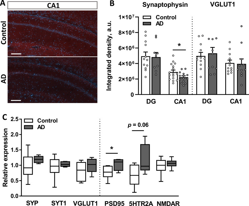
Fig. 8.
The i.c.v. injection of AβOs alters synaptic plasticity in the hippocampus. A Representative images of immunofluorescence staining for synaptophysin within the CA1 and DG of AD and control rats. Scale bar, 200 µm. B Quantification of synaptophysin- and VGLUT1-positive IR areas in the DG and CA1 regions of the hippocampus, which are reported as integrated densities. Data are from two–three sections per rat, 6 rats per group. C The relative mRNA expression of synaptic markers measured using qPCR. Data were normalized to two reference genes: ACTB and RPL13A. B–C Data are presented as the means ± SEM. *p < 0.05. n = 6 rats per group

