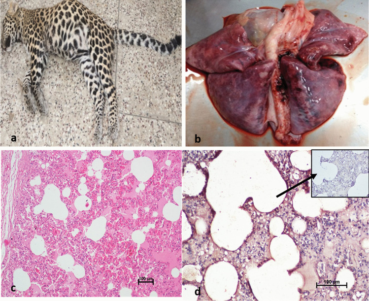
Fig. 2.
Lung from necropsied leopard showing severe inflammatory changes. a Carcass of leopard cub. b Representative macroscopic image of affected lung of animal. c Representative microscopic image (objective × 20) of section of lungs fixed in 10% formalin and stained with HE, showing diffuse inflammatory changes like hemorrhages and congestion and thickening of alveolar septum. d Representative microscopic image showing strong immune reactivity to SARS-COV-2 in the alveolar septal cells and inflammatory cells. IHC DAB × 200. Inset: antibody control, IHC DAB × 20

