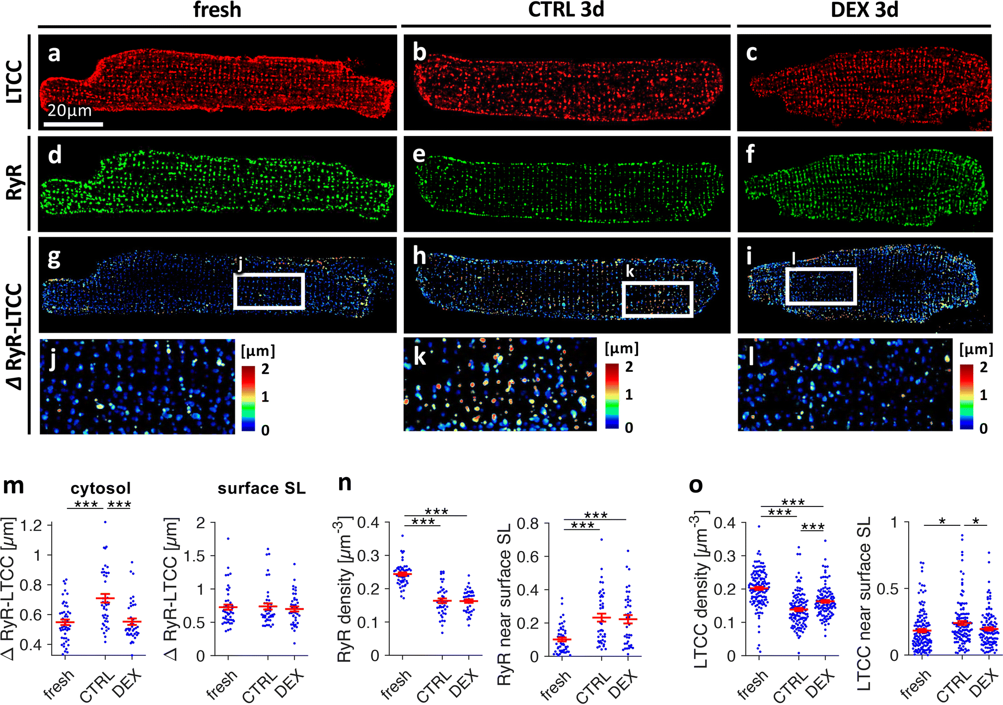
Figure 4.

Effect of dexamethasone on distributions and spatial relationship of L-type Ca2+ channels (LTCC) and ryanodine receptors (RyR) in cultured rat cardiomyocytes. Freshly isolated myocytes (fresh) and myocytes cultured for 3d in vehicle (CTRL3d) or 1μmol/L dexamethasone (DEX3d) were fixed, co-immunostained for LTCC and RyR and analysed by 3D confocal microscopy. A-C, LTCC, D-F, RyR, G-L, color-coded distances between RyR clusters and their closest LTCC cluster (ΔRyR-LTCC). M, ΔRyR-LTCC in the cytosol (≥ 0.5μm from the surface sarcolemma, SL) and ΔRyR-LTCC close (< 0.5μm) to the surface SL. N, RyR cluster density (clusters per μm3) and fraction of RyR signal detected close (< 0.5μm) to the surface SL. O, LTCC cluster density and fraction of LTCC signal detected close to the surface SL. Scale bar in A also applies to B-I. ***p<0.001, **p<0.01, *p<0.05, n (cells/animals) = 45/10, 43/7, 40/7 in fresh, CTRL3d, DEX3d, respectively. Two-tailed Welch’s t-test was used for all tests, with multiple comparison correction.
