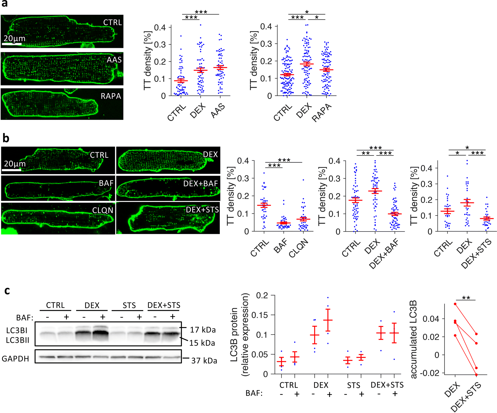
Figure 8.

Effects of autophagy enhancers and blockers on t-system density in isolated rat cardiomyocytes. A, Confocal images and quantification of t-tubule (TT) skeleton density of Di8-ANEPPS-stained myocytes after 3 days in culture, treated with vehicle (CTRL), amino-acid free medium (amino acid starvation, AAS), or 1μmol/L rapamycin (RAPA). n (cells/animals) = 38/5, 40/5, 38/5 in CTRL, DEX, AAS, respectively, and 126/14, 97/14, 97/14 in CTRL, DEX, RAPA, respectively. B, Confocal images and quantification of TT density of myocytes after 2 days in culture, treated with vehicle (CTRL), 100nmol/L bafilomycin A1 (BAF), 5μmol/L chloroquine (CLQN), 1μmol/L dexamethasone (DEX), DEX + 100nmol/L bafilomycin A1 (DEX+BAF) or DEX + 10nmol/L staurosporine (DEX+STS). n (cells/animals) = 40/5, 40/5, 40/5 in CTRL, BAF, CLQN, respectively, and 60/8, 61/8, 61/8 in CTRL, DEX, DEX+BAF, respectively, and 30/4, 31/4, 29/4 in CTRL, DEX, DEX+STS, respectively. Example images were chosen to be representative of mean TT densities. ***p<0.001, **p<0.05 (Welch’s t-test with multiple comparison correction) C, Example and quantitative analysis of LC3BII Western blots from myocytes cultured for 1d (CTRL, DEX, 10 nmol/L staurosporine (STS), DEX+STS) that were treated with either 100nmol/L bafilomycin A1 (BAF+) or with vehicle (BAF−) for 2.5 h before freezing, to assess LC3BII accumulation as a measure of autophagic flux. Accumulated LC3BII due to BAF treatment was calculated by pairwise subtraction of BAF− from BAF+ LC3BII protein levels from n = 4 matched cell isolations. **p<0.01 (paired t-test)
