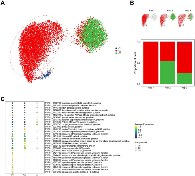
Fig 3. Clustering and differential gene expression analysis of P. vivax sporozoites.
(A) UMAP of integrated P. vivax sporozoite transcriptomes coloured by cluster (Leiden algorithm; resolution parameter = 0.1). (B) UMAP of integrated P. vivax sporozoite transcriptomes split by replicate (upper) and the percentage of sporozoites in each cluster from the replicate (lower). (C) Dot plot showing top markers that distinguish each of the three clusters. The size of the dot corresponds to the percentage of sporozoites expressing the gene. Scale bar: normalised expression, scaled.

