. 2022 Apr 11;21(6):999–1009. doi: 10.1158/1535-7163.MCT-21-0726

Figure 1.

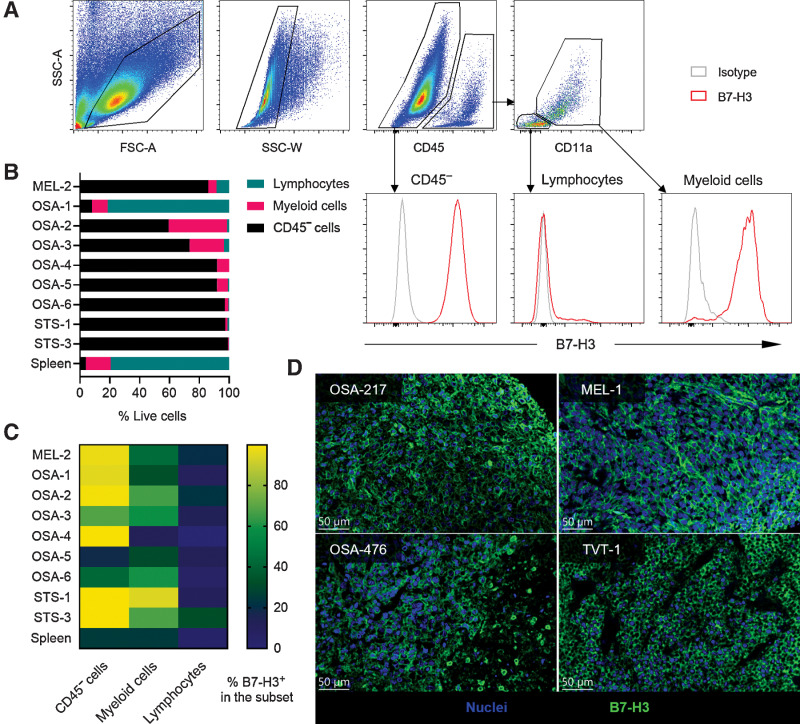
Figure 1. B7-H3 is expressed on canine solid tumors. A, Gating strategy of CD45- cells, tumor-infiltrating lymphocytes, and myeloid cells on a STS sample (STS-1), and the expression levels of B7-H3. B, Summary of the frequencies of CD45- cells, lymphocytes, and myeloid cells from each sample. Spleen samples were included as a negative control. C, Expression level of B7-H3 on different cell subsets from each tumor sample. Tumor cells generally have the highest level of B7-H3, followed by myeloid cells. D, Representative IHC results of surface B7-H3 expression on canine osteosarcoma, melanoma (MEL) and TVT. Blue indicates nuclei and green indicates B7-H3 staining.

B7-H3 is expressed on canine solid tumors. A, Gating strategy of CD45 cells, tumor-infiltrating lymphocytes, and myeloid cells on a STS sample (STS-1), and the expression levels of B7-H3. B, Summary of the frequencies of CD45 cells, lymphocytes, and myeloid cells from each sample. Spleen samples were included as a negative control. C, Expression level of B7-H3 on different cell subsets from each tumor sample. Tumor cells generally have the highest level of B7-H3, followed by myeloid cells. D, Representative IHC results of surface B7-H3 expression on canine osteosarcoma, melanoma (MEL) and TVT. Blue indicates nuclei and green indicates B7-H3 staining.