. 2022 Apr 1;21(6):974–985. doi: 10.1158/1535-7163.MCT-21-0599

Figure 2.

Figure 2. Selection of LY6G6D and CD3 arms for the optimized LY6G6D-TDB. A, Dose–response analysis of HT55 cell killing mediated by TDBs of anti-LY6G6D clones from different epitope bins paired with the low-affinity CD3 arm. Cell killing was measured by CellTiter-Glo reagent at 48-hour time point. B and C, CD8+ and CD4+ T-cell activation induced by TDBs of anti-LY6G6D clones from different epitope bins. T-cell activation was measured at 48-hour time point by flow cytometry. D, Killing of HT55 cells by 1G4-TDB paired with high- or low-affinity CD3 arm. 48-hour time point. EC50 values were 3.31 ± 1.91 ng/mL and 86.00 ± 60.61 ng/mL from 4 PBMC donors, respectively. E and F, CD8+ and CD4+ T-cell activation induced by 1G4-TDB paired with high- or low-affinity CD3 arm. A–C, The representative data of 3 PBMC donors. D–F, The representative data of 4 PBMC donors. Cell killing data are shown as means ± SD of triplicate wells.

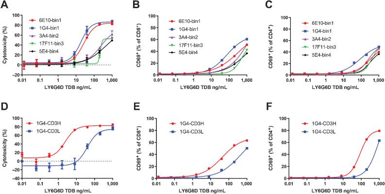
Selection of LY6G6D and CD3 arms for the optimized LY6G6D-TDB. A, Dose–response analysis of HT55 cell killing mediated by TDBs of anti-LY6G6D clones from different epitope bins paired with the low-affinity CD3 arm. Cell killing was measured by CellTiter-Glo reagent at 48-hour time point. B and C, CD8+ and CD4+ T-cell activation induced by TDBs of anti-LY6G6D clones from different epitope bins. T-cell activation was measured at 48-hour time point by flow cytometry. D, Killing of HT55 cells by 1G4-TDB paired with high- or low-affinity CD3 arm. 48-hour time point. EC50 values were 3.31 ± 1.91 ng/mL and 86.00 ± 60.61 ng/mL from 4 PBMC donors, respectively. E and F, CD8+ and CD4+ T-cell activation induced by 1G4-TDB paired with high- or low-affinity CD3 arm. A–C, The representative data of 3 PBMC donors. D–F, The representative data of 4 PBMC donors. Cell killing data are shown as means ± SD of triplicate wells.