
Figure 1.
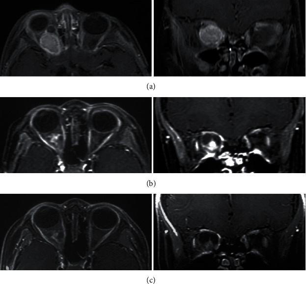
Contrast-enhanced MRI of pediatric OPG before and after single fraction GKS (16.0 Gy). A male OPG patients aged 30 months was observed with proptosis of the right eye, accompanied with VA damage for more than 2 months. (a) MRI at GKS. The tumor is located in the right orbit and surrounded the optic nerve and was homogeneous enhanced on MRI. The baseline VA was 0.15 on the right. (b) Three years after GKS, the tumor volume was smaller than baseline, there was intratumoral necrosis and cyst formation, and tumor enhancement effect declined. The VA remained 0.15, and the proptosis improved significantly. (c) Five years after GKS, the tumor volume remained stable, and tumor enhancement effect declined continuously. The VA was less than 0.1.