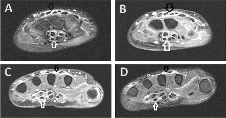
Fig. 1.
Axial MR image of the right wrist demonstrated T2 hyperintense signal (A) with T1 post-contrast enhancement (B) throughout the flexor (white arrow) and extensor (black arrow) tendon sheaths at the level of the carpal tunnel (white arrow). These findings were also seen at the level of the metacarpals on T2 (C) and T1 post-contrast (D) MR images. Findings were bilateral

