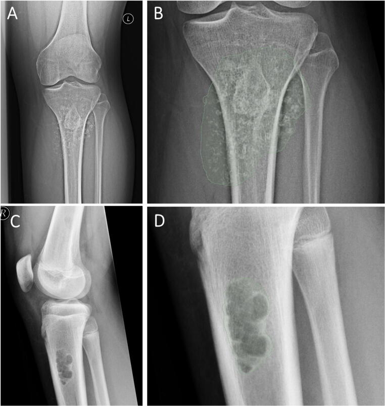
Fig. 5.
A and B Example of a malignant tumor in the tibia of a 33-year-old male with a chondrosarcoma. A shows the radiograph and B shows the segmentation for the radiomics extraction. The artificial neural network model combining both demographic and radiomic information correctly predicted a malignant tumor with a certainty of 86%. C and D Example of a benign tumor in the proximal tibia of a 15-year-old male with a non-ossifying fibroma. A shows the radiograph and B shows the segmentation for the radiomics extraction. The artificial neural network model using the combination of both, the demographic and radiomic information, correctly predicted a benign tumor with a certainty of 93%

