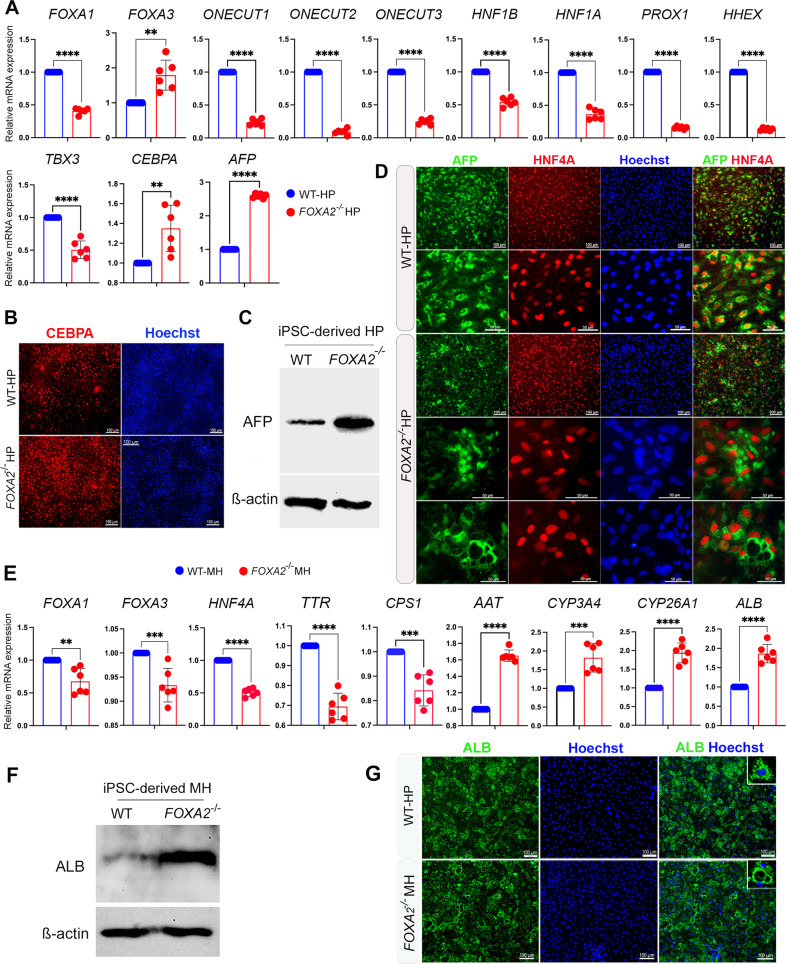
Fig. 2. Effect of FOXA2 knockout on iPSC-derived hepatocytes.
A RT-qPCR analysis showing the mRNA expression of hepatic progenitor (HP) markers, FOXA1, FOXA3, ONECUT1, ONECUT2, ONECUT3, HNF1B, HNF1A, PROX1, HHEX, TBX3, CEBPA, and AFP in FOXA2−/− HP relative to wild type (WT) controls (n = 6). B Immunofluorescence images showing the expression of CEBPA in HP derived from WT-iPSCs and FOXA2−/−iPSCs. C Western blot analysis showing the upregulation of AFP in HP lacking FOXA2 compared to WT controls. D Immunofluorescence analysis showing co-expression of AFP and HNF4A in HP derived from WT-iPSCs and FOXA2−/−iPSCs. Note the pattern of AFP expression in FOXA2−/−HP. E RT-qPCR analysis showing the mRNA expression of mature hepatocyte (MH) markers, FOXA1, FOXA3, HNF4A, AAT, TTR, CPS1, CYP3A4, CYP26A1, and ALB in FOXA2−/− MH relative to WT controls (n = 6). F Western blotting showing the upregulation of ALB protein in FOXA2−/− MH compared to WT. G Immunofluorescence showing ALB protein expression in FOXA2−/−MH compared to WT controls. Note the ALB distribution around cytoplasmic vacuoles. Nuclei were counterstained with Hoechst. The data are presented as mean ± SD. *p < 0.05, **p < 0.01, ***p < 0.001.

