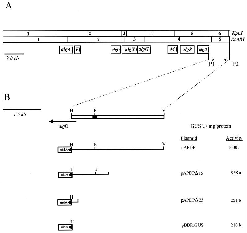
FIG. 1.
(A) Physical and functional map of the alginate structural gene cluster in Pseudomonas syringae pv. syringae FF5. The arrows within each open reading frame indicate the direction of translation. The locations of the primers (P1 and P2) used to amplify the algD promoter region are indicated. Abbreviations: F, algF; 44, alg44. (B) Expanded view of the region amplified with primers P1 and P2. The location and orientation of the coding region for algD are shown (horizontal arrow). The black boxes flanking the EcoRI site indicate the consensus sequence recognized by AlgT (ς22). The location and orientation of the algD::uidA transcriptional fusions are indicated; GUS activity is shown in the column adjacent to each construct. Values followed by the same letter were not significantly different (P = 0.01). Abbreviations: E, EcoRI; H, HindIII; V, EcoRV.

