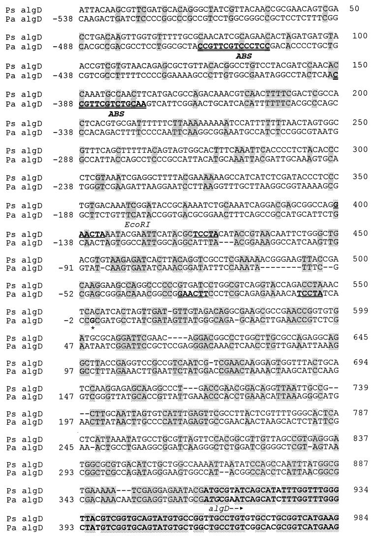
FIG. 3.
Alignment of the algD promoter sequences from P. syringae pv. syringae FF5 (Ps algD) and P. aeruginosa (Pa algD). The P. aeruginosa algD promoter was previously reported (24, 39); the nucleotides for this sequence are shown on the left, with +1 (asterisk) corresponding to the transcriptional start site. Nucleotides for the P. syringae pv. syringae algD promoter are shown on the right. The EcoRI site in the P. syringae sequence corresponds to the left border of EcoRI fragment 5 in Fig. 1A. Gaps (––) were used to maximize the alignment, and identical bases are shaded. The AlgR1 binding sites (ABS) in the P. aeruginosa algD promoter are shown in bold and double-underlined. The ς22 recognition sequence in both species is indicated in bold and single-underlined. The algD translational start site and coding region are shown in bold (algD–→).

