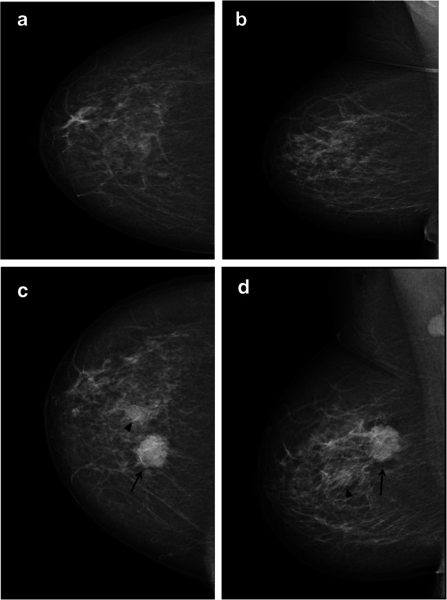
Fig. 5.
The craniocaudal and mediolateral oblique mammograms of the right breast at index (a and b) and subsequent screening (c and d) from a 54-year-old woman diagnosed with subsequent screen-detected cancer after false-positive index screening. The examination was characterized as a one-plane asymmetry in the craniocaudal view at index screening. At subsequent screening, a circumscribed mass in the upper medial quadrant (arrow) and a smaller mass, located more lateral and inferior (arrowhead), were both histologically verified as cancers

