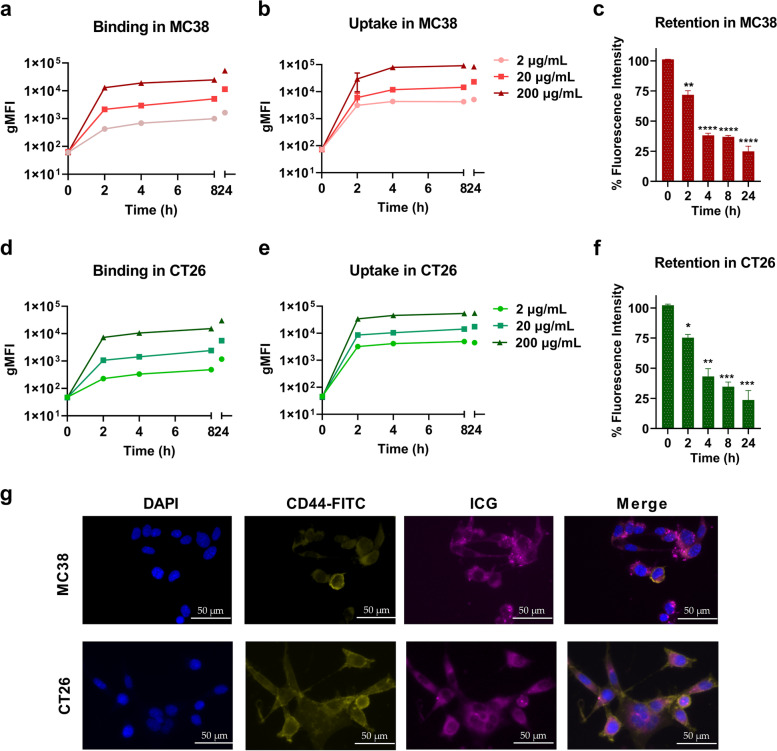
Fig. 1.
Cellular properties of the photosensitizer ICG in vitro (a) Cellular binding and (b) uptake assays with 2 µg/mL, 20 µg/mL, and 200 µg/mL ICG in MC38 cells over time by incubating cells with ICG at 4 °C and 37 °C, respectively. Detection was performed by flow cytometry and represented as gMFI of ICG. (c) Retention of ICG (50 µg/mL) in MC38 cells after 4 h co-incubation, collected cells were washed and detected by flow cytometry. (d) Cellular binding and (e) uptake assays with 2 µg/mL, 20 µg/mL, and 200 µg/mL ICG in CT26 cells over time by incubating cells with ICG at 37 °C and 4 °C, respectively. (f) Retention of ICG (50 µg/mL) in CT26 cells after 4 h co-incubation, collected cells were washed and detected by flow cytometry. The fluorescence signal positive population is shown as a percentage of CRC cells. (g) Fluorescence microscopy images of ICG (50 µg/mL)-treated MC38 and CT26 cells after 4 h of incubation. Scale bar = 50 μm

