FIGURE 3.

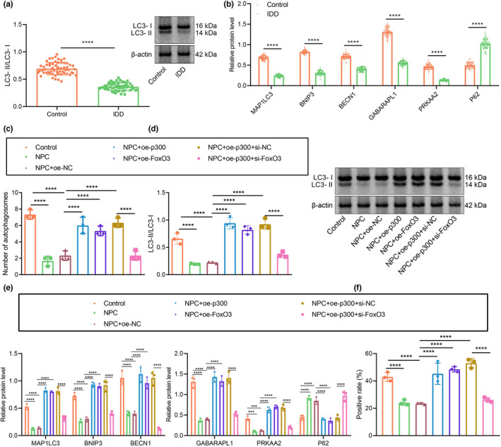
p300 promotes FOXO3 expression and enhances autophagy of NPCs. (a) The ratio of LC3‐II/LC3‐I in clinical samples of healthy controls and patients with IDD as determined by Western blot assay. n = 58. (b) The protein expression of autophagy‐related factors in clinical samples of healthy controls and patients with IDD as determined by Western blot assay. (c) Observation of autophagy in human NPCs by TEM. (d) The ratio of LC3‐II/LC3‐I in NPCs as determined by Western blot assay. (e) The protein expression of autophagy‐related factors in NPCs as determined by Western blot assay. (f) Autophagic flux determined by mRFP‐GFP‐LC3 assay. The expression and location of LC3 in NPCs as examined by immunofluorescence staining. The measurement data were expressed as mean ± standard deviation. * p < 0.05. ** p < 0.01. *** p < 0.001. **** p < 0.0001. Comparison between two groups was conducted using independent sample t‐test. Comparison among multiple groups was conducted using one‐way ANOVA, followed by Tukey post‐hoc test
