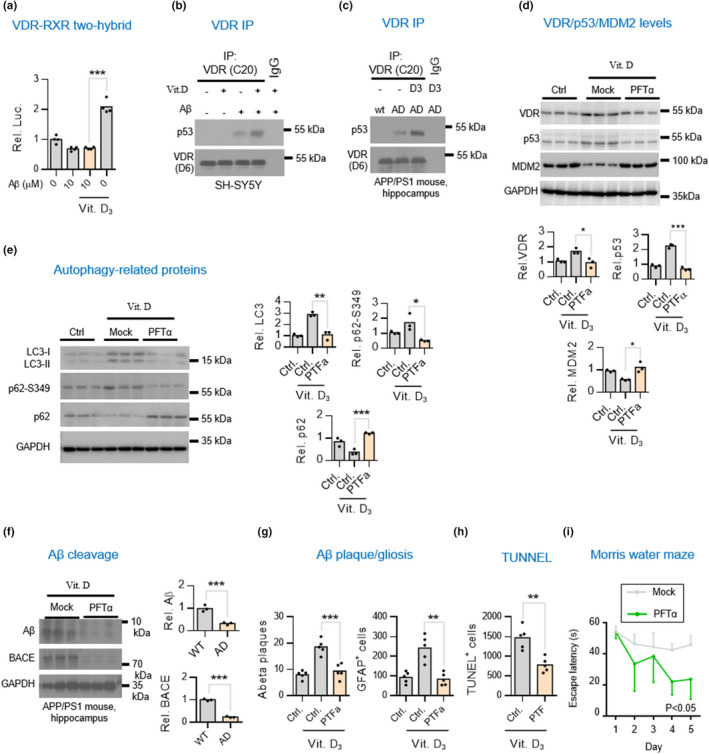
FIGURE 2.

Vitamin D supplementation enhances VDR/p53 but not VDR/RXR complex in worsening brain pathology in APP/PS1 AD mice. (a) Mammalian two‐hybrid assays for studies of the interaction of VDR with RXR in neuronal cells exposed to Αβ plus with vitamin D3. SH‐SY5Y cells were treated Aβ42 for 6 h and then co‐treated with 10 nM calcitriol for additional 6 h prior to harvesting for mammalian two‐hybrid luciferase assays. (b,c) Western blot analysis of co‐immunoprecipitation of VDR/p53 complex in SH‐SY5Y cells and hippocampal tissues of APP/PS1 mice. (d) Western blot analysis of VDR, p53, and MDM2 in the hippocampal lysates of APP/PS1 mice treated with or without p53 inhibitor. 4.5‐month‐old APP/PS1 mice raised on vitamin D3‐sufficient diets were intraperitoneally injected weekly with 3 mg/kg of p53 inhibitor pifithrin‐α (PFTα) for 7.5 months before harvesting hippocampal tissues for analysis. Densitometrical quantification of VDR, p53, and MDM2 bands were normalized to GAPDH (lower panel). *p < 0.05; **p < 0.01; ***p < 0.001 by unpaired t‐test. (e) Western blot analysis of autophagic markers LC3, p62, and ser349 phosphorylated p62 (p62‐S349) in the hippocampal lysates of APP/PS1 mice injected with or without PFTα. Densitometrical quantification of LC3, p62‐S349, and p62 bands were normalized to GAPDH (right panel). (f) Western blot analysis of Αβ and BACE levels in the hippocampal lysates of APP/PS1 mice injected with or without PFTα. Densitometrical quantification of Aβ and BACE bands were normalized to GAPDH (right panel). (g,h) p53 inhibitor amelioration of vitamin D3‐aggravated Aβ aggregation and apoptosis. The Aβ, GFAP, and TUNEL‐positive signals in five consecutive sections per animal (n = 5) was quantified by ImageJ and presented as the mean ± SD. Scale bars, 50 μm. (i) Cognitive performance assays for the AD mice treated with p53 inhibitor. APP/PS1 mice were given with or without weekly injections of PTFα (n = 6 mice) starting at the age of 4.5‐month. APP/PS1 mice at 12‐month of age were used for the Morris Water Maze test
