
FIGURE 2.
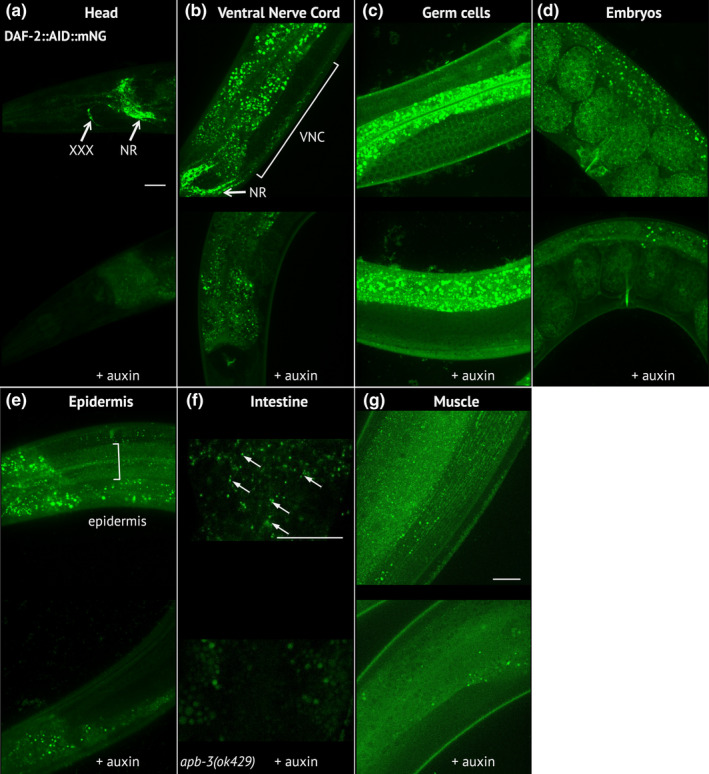
DAF‐2::AID::mNG is effectively downregulated in the presence of ubiquitously expressed TIR1 after auxin treatment. (a–g) Images of DAF‐2::AID::mNG in 1‐day‐old daf‐2(kr462) adults expressing ubiquitous TIR1 and grown in the absence of auxin (upper panels) or after 24 h of auxin treatment (lower panels). Images focus on specific body regions: the head (a), showing strong expression in the nerve ring (NR) and the XXX cells; the neuronal cell bodies of the ventral nerve cord (VNC) (b); the proliferating germ cells (c); the embryos (d); the epidermal syncytium (e); the intestine (f) and the body wall muscles (g). For the intestine (f), images were taken in apb‐3(ok429) mutant background in order to reduce unspecific intestinal autofluorescence (arrows indicate the specific DAF‐2::AID::mNG associated signal). In all images, the remaining staining of the gut after auxin treatment corresponds to nonspecific autofluorescence that varies between animals. Similar results were obtained in 7‐day‐old animals (data not shown). Scale bars: 20 μm