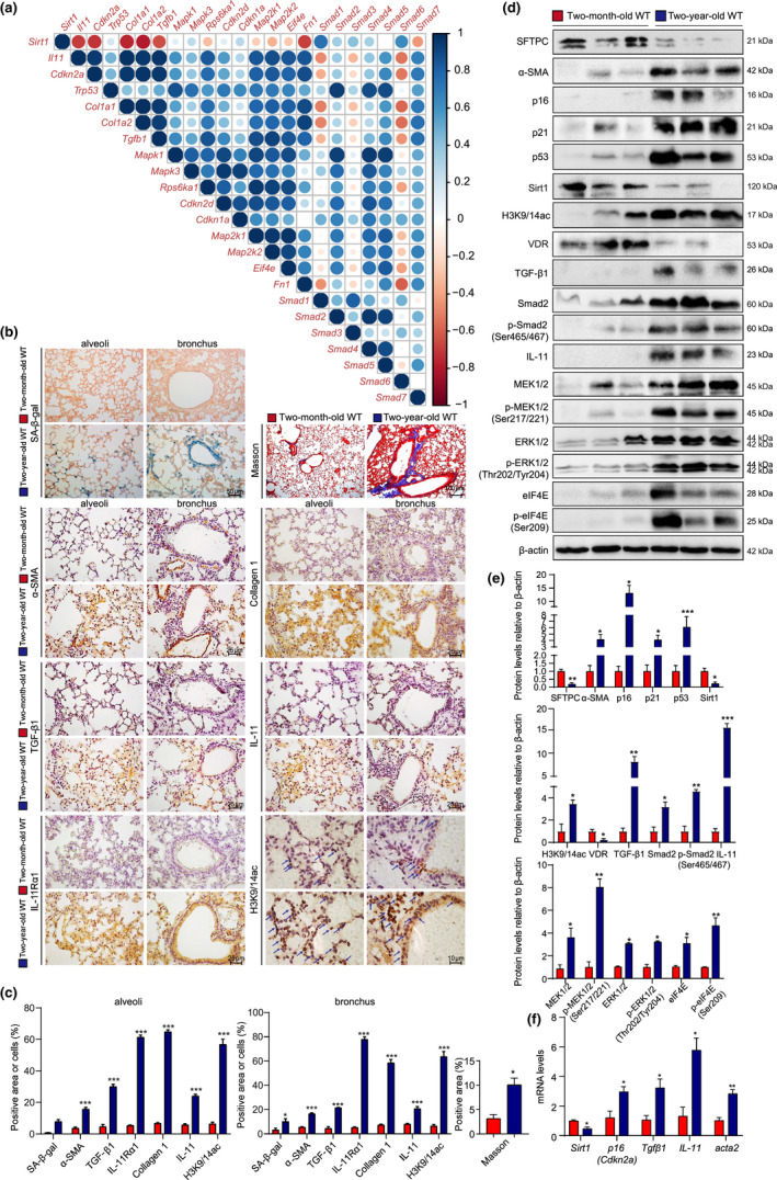
FIGURE 1.

Pulmonary Sirt1 and serum VD decreases with physiological aging, activating TIME signaling, and promoting SAPF. (a) Gene correlation analysis using RNAseq data of pulmonary GFP+CD45−CD31−EpCAM− fibroblasts from physiologically aged (18 months old) Col1α1‐GFP mice treated with or without bleomycin. The lungs from young (2 months old) and physiologically aged (2 years old) WT mice were detected. (b) Representative micrographs of paraffin‐embedded pulmonary tissue stained histochemically for SA‐β‐gal and with Masson's trichrome (Masson), immunohistochemically for α‐SMA, collagen 1, TGF‐β1, IL‐11, IL‐11Rα1, and H3K9/14ac in alveoli and bronchus, with hematoxylin staining the nuclei. (c) Alveolar and bronchial areas positive for Masson staining, α‐SMA, collagen 1, TGF‐β1, IL‐11, IL‐11Rα1, or H3K9/14ac. (d) Western blotting of pulmonary extracts showing SFTPC, α‐SMA, p16, p21, p53, Sirt1, H3K9/14ac, VDR, TGF‐β1, Smad2, p‐Smad2(Ser465/467), IL‐11, MEK1/2, p‐MEK1/2(Ser217/221), ERK1/2, p‐ERK1/2(Thr202/Tyr204), elF4E, and p‐elF4E(Ser209). β‐actin was used as the loading control. (e) Protein levels relative to β‐actin were assessed by densitometric analysis. (f) Sirt1, p16, Tgfβ1, IL‐11, and acta2 mRNA levels in lungs of young and aged mice by real‐time RT‐PCR, calculated as ratio to β‐actin mRNA and expressed relative to control. Six mice per group were used for experiments. Values are the mean ± SEM of three determinations per group. *p < 0.05, **p < 0.01, ***p < 0.001 compared with the young mice
