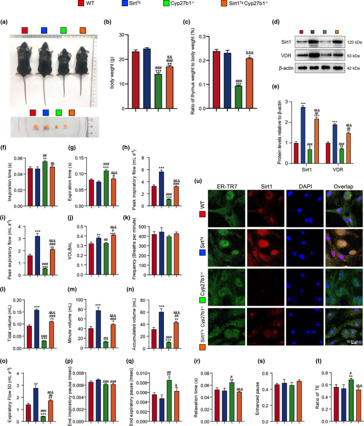
FIGURE 2.

Sirt1 overexpression improves pulmonary dysfunction in VD‐deficient mice. (a) Representative appearances of 9‐week‐old WT, Sirt1 Tg , Cyp27b1 −/− and Sirt1 Tg Cyp27b1 −/− mice and whole view of the thymus. (b, c) Body weight of different genotyped mice and the ratio of thymus weight relative to body weight. (d, e) Western blotting of pulmonary extracts showing Sirt1 and VDR protein levels. Pulmonary function was detected by the whole‐body plethysmography for (f) inspiration time (s), (g) expiration time (s), (h) peak inspiratory flow (mL s−1), (i) peak expiratory flow (mL s−1), (j) VOLBAL, (k) frequency (breaths per minute), (l) tidal volume (mL), (m) minute volume (mL), (n) accumulated volume (mL), (o) expiratory flow 50 (mL s−1), (p) end inspiratory pause (ms), (q) end expiratory pause (ms), (r) relaxation time (s), (s) enhanced pause, and (t) ratio of TE. Six mice per group were used for experiments. (u) Representative micrographs of cells immunofluorescently stained for Sirt1 and ER‐TR7, with DAPI staining the nucleus. Values are the means ± SEM of six determinations per group. *p < 0.05, **p < 0.01, ***p < 0.001 compared with the WT group; # p < 0.05, ## p < 0.01, ### p < 0.001 compared with the Sirt1 Tg group; & p < 0.05, && p < 0.01, &&& p < 0.001 compared with Cyp27b1 −/− group
