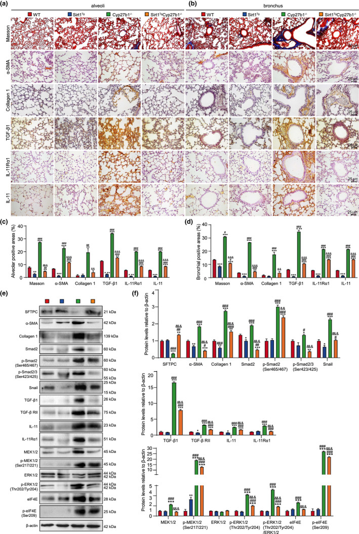
FIGURE 3.

Sirt1 overexpression improves PF through inhibiting TIME signaling in VD‐deficient mice. (a, b) Representative micrographs of paraffin‐embedded pulmonary stained histochemically for Masson's trichrome (Masson), immunohistochemically for α‐SMA, collagen 1, TGF‐β1, IL‐11Rα1, and IL‐11 in alveoli (a) and bronchus (b), with hematoxylin staining the nuclei. Percentage of alveolar (c) and bronchial (d) areas positive for Masson staining, α‐SMA, Collagen 1, TGF‐β1, IL‐11Rα1, or IL‐11. (e) Western blotting of pulmonary extracts showing SFTPC, α‐SMA, Collagen 1, Smad2, p‐Smad2(Ser465/467), p‐Smad2/3(Ser423/425), Snail, TGF‐β1, TGF‐β RII, IL‐11, IL‐11Rα1, MEK1/2, p‐MEK1/2(Ser217/221), ERK1/2, p‐ERK1/2(Thr202/Tyr204), elF4E, and p‐elF4E(Ser209). β‐actin was used as the loading control. (f) Protein expression relative to β‐actin was assessed by densitometric analysis. Six mice per group were used for experiments. Values are the mean ± SEM of six determinations per group. *p < 0.05, **p < 0.01, ***p < 0.001 compared with the WT group; # p < 0.05, ## p < 0.01, ### p < 0.001 compared with the Sirt1 Tg group; && p < 0.01, &&& p < 0.001 compared with the Cyp27b1 −/− group
