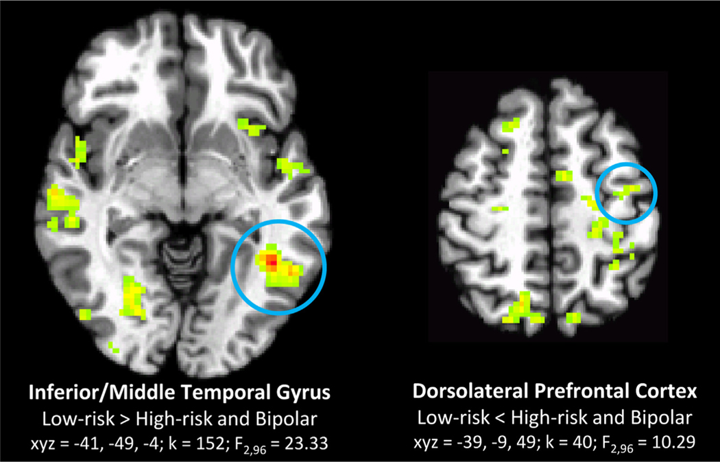
FIGURE 2.
Candidate risk endophenotypes: regions where activation related to face emotion labeling in low-risk (LR) youth differ from those of high-risk (HR) youth and youth with bipolar disorder (BD), across all stimuli (group main effect). Note: Circled clusters correspond to statistical information below brain image; other clusters are significant group differences that do not fit the pattern of a risk endophenotype (i.e., LR≠HR+BP). Axial sections shown in radiological view (left = right) in all figures. Clusters for all figures significant at whole-brain corrected p < .05.

