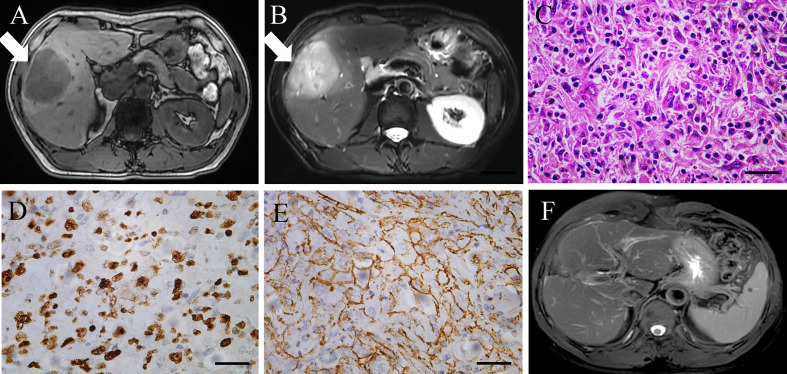
Figure 1.
(A, B) Enhanced MRI scan of the liver lesion before surgery. (C–E) Histopathological finding (hematoxylin and eosin staining, Ki67, and β-catenin), poorly differentiated intrahepatic sarcomatoid cholangiocarcinoma. Magnification, ×400. Scale bar: 50 µm. (F) MRI scan of the liver lesion after surgery in February 2022.

