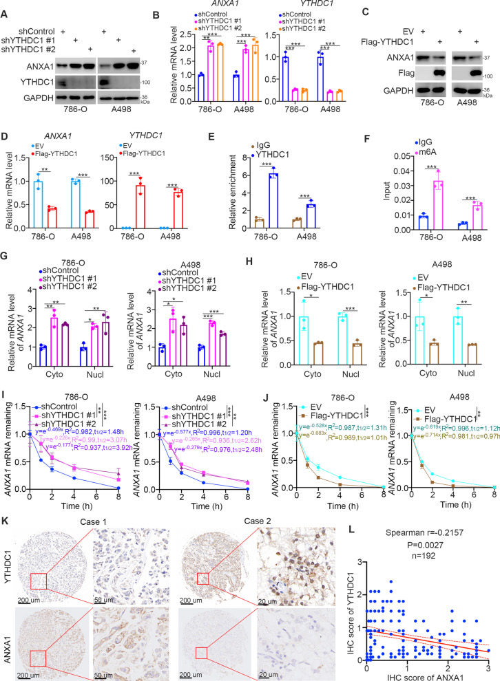
Fig. 4.
YTHDC1 represses ANXA1 expression in ccRCC cells. A and B, 786-O and A498 cells were transfected with the indicates shRANs for 72 h. Cell were harvested for western blot analysis and RT-qPCR analysis. Data presents as mean ± SD with four replicates. **, P < 0.01; ***, P < 0.001. C and D, 786-O and A498 cells were transfected with the indicates plasmids for 24 h. Cell were harvested for western blot analysis and RT-qPCR analysis. Data presents as mean ± SD with four replicates. **, P < 0.01; ***, P < 0.001. E, The IgG or YTHDC1 antibodies was used to performed the ChIP-qPCR assay in 786-O and A498 cells. Data presents as mean ± SD with four replicates. ***, P < 0.001. F, Magna m6A MeRIP kit was used to performed MeRIP-qPCR assay in 786-O and A498 cells. Data presents as mean ± SD with four replicates. **, P < 0.001. G, 786-O and A498 cells were transfected with indicated shRNAs for 72 h. Cells were harvested, and the RNA was extracted from the cytoplasm (cyto) or nucleus (mucl) respectively. The RT-qPCR assay was used to detect the mRNA of ANXA1 in 786-O and A498 cell. Data presents as mean ± SD with four replicates. *, P < 0.05; **, P < 0.01; ***, P < 0.001. H, 786-O and A498 cells were transfected with indicated plasmids for 24 h. Cells were harvested, and the RNA was extracted from the cytoplasm (cyto) or nucleus (mucl) respectively. The RT-qPCR assay was used to detect the mRNA of ANXA1 in 786-O and A498 cell. Data presents as mean ± SD with four replicates. *, P < 0.05; **, P < 0.01; ***, P < 0.001. I, 786-O and A498 cells were transfected with indicated shRNAs for 72 h. Then, cells treated with actinomycin D (5 µg/mL). Then, cells were collected at the different time points. Total RNAs were extracted and analyzed by RT-qPCR. The mRNA expression for each group was normalized to β-actin. J, 786-O and A498 cells were transfected with indicated plasmids for 72 h. Then, cells treated with actinomycin D (5 µg/mL). Then, cells were collected at the different time points. Total RNAs were extracted and analyzed by RT-qPCR. The mRNA expression for each group was normalized to β-actin. K and L, The IHC staining was performed in the tissue microarray of renal cancer by using the YTHDC1 and ANXA1 antibodies. The typical image was shown in panel K. The correlation between ANXA1 and YTHDC1 was shown in panel L, P = 0.0027

