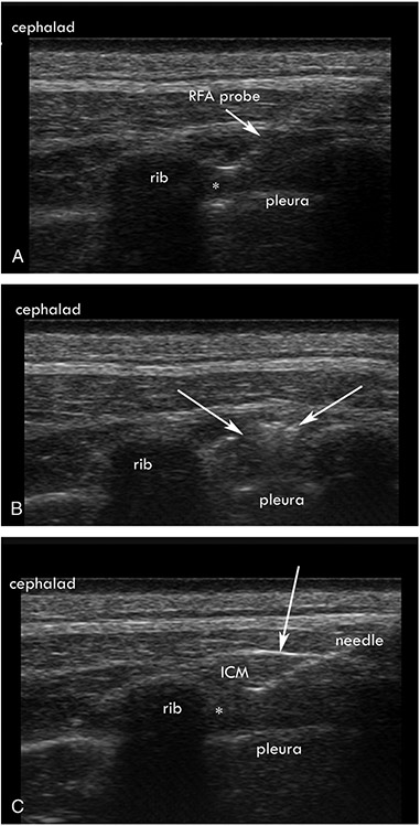
FIGURE 1.
A, Ultrasound-guided radiofrequency ablation (RFA) showing placement of the tip (*) at the inferior border of the corresponding rib within the internal intercostal muscle (ICM) and adjacent to innermost intercostal muscle. B, Ultrasound image of the muscle after RFA, with the arrows showing hyperechoic changes in the ICM. C, Ultrasound-guided alcohol neurolysis, with a 25-gauge needle (*) showing the location of the needle tip and injection of local anesthetic. Arrows point to the tenting of the ICM, indicating spread of the alcohol into the ICM and innermost intercostal muscle.

