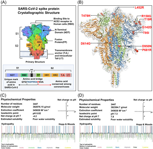
Figure 1.

A comparative analysis to determine the impact of targeted mutations on the physiochemical properties of S protein. (A) Crystallographic conformation of the SARS‐CoV‐2 S protein (B) Localization of the targeted nine missense mutations found in the delta variant. The three amino acid chains of the trimeric S protein were encoded by three different colors, blue, green, and orange. The mutations were marked by red ball‐stick shape. (C, D) Physiochemical properties of both of the wild and mutated S protein. Top layer of the Hopp & Wood hydropathy plot remarked hydrophilic amino acids and the bottom layer indicated the hydrophobic amino acids. SARS‐CoV‐2, severe acute respiratory syndrome coronavirus 2
