
Fig. 1. Loss of IL-6 reduces cartilage degradation in male mice with PTOA.
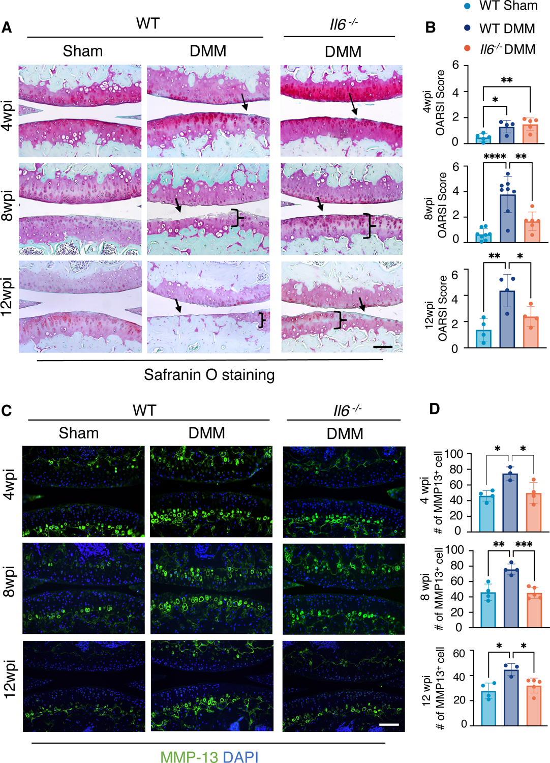
(A and B) Safranin-O staining of knee joint sections (A) and OARSI scores quantifying the cartilage phenotypes (B) in WT and Il6−/− male mice at 4, 8, and 12 weeks after sham or DMM surgery. N ≥ 5 mice per condition (4wpi); N≥ 6 mice per condition (8wpi); N≥ 4 mice per condition (12wpi). wpi, weeks post-injury. Scale bar, 100 μm. (C) Immunofluorescence staining for MMP-13 in knee sections from WT or Il6−/− male mice at 4, 8, and 12 weeks after sham or DMM surgery. DAPI indicates nuclei. Scale bar, 100 μm. (D) Quantification of the number of cells with MMP-13 staining. Green fluorescence encircling cells was used to identify individual MMP-13+ chondrocytes. N≥ 3 mice per condition. One-way ANOVA. All quantitative data represent mean ± SD. * p<0.05, ** ≤p<0.01, *** ≤p<0.001, **** p<0.0001.