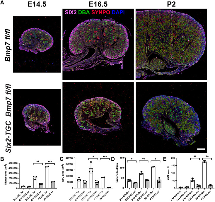
Fig. 1.
NPCs and ureteric bud branching in Bmp7 mutant kidneys. (A) Time points at top of each column, genotypes at left of each row. Pink, Six2; green, DBA lectin; red, synaptopodin. The olive-green color in the E16.5 and P2 panels is autofluorescence characteristic of kidney tubules in paraffin-embedded tissues. Scale bar: 250 µm. (B) Cross-sectional kidney area measured at equivalent sections through the papilla at the widest diameter of the kidney. (C) NPC area measured by AF647 fluorescence. (D) Ureteric bud tips measured by counting green-stained tips within clusters of NPCs. (E) Glomerular number obtained by counting structures stained for synaptopodin. Data are mean±s.d. *P<0.05, **P<0.01, ***P<0.001 (unpaired two-tailed t-test). Three biological replicate kidneys were examined at each time point.

