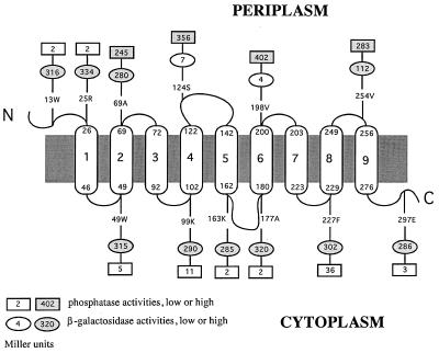
FIG. 2.
Experimentally derived model for the structure of the MntB protein with nine transmembrane domains (numbered capsules) and locations of alkaline phosphatase and β-galactosidase fusions (rectangles and ovals, respectively). The small numbers at the tops and bottoms of the capsules correspond to positions of amino acid residues in MntB. Sites of fusions are indicated by similar numbers followed by the one-letter designations of the corresponding amino acids.

