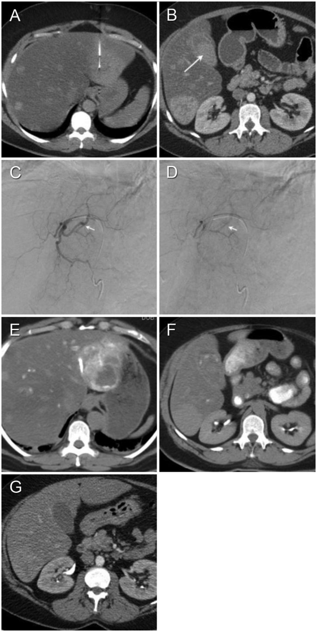
Figure 3.

A 51-year-old female with presents with multiple liver lesions (Patient #10 in Tables 1a–c). (A) Non-enhanced interventional CT shows target lesion and biopsy needle vector and tip. (B) Contrast-enhanced axial CT on day 4 post-biopsy to evaluate severe right upper quadrant pain demonstrates hyperdense gallbladder contents compatible with hemorrhage. (C, D) Diagnostic arteriogram performed on day 14 post-biopsy for severe persistent pain shows arterio-venous fistula without active extravasation. The left hepatic artery was embolized to stasis with 6 cc 100–300 μm embospheres and 1 cc 100 μm PVA (not shown). (E, F) Non-enhanced post-embolization CT hyperdensity of the tumor (indicating successful embolization of the tumor) as well as the lumen of the gallbladder, duodenum and stomach (indicating hemobilia with enterogastric reflux). (G) Contrast-enhanced axial CT performed 21 days post-biopsy shows interval evolution and near-complete resolution of subcapsular hematoma.
