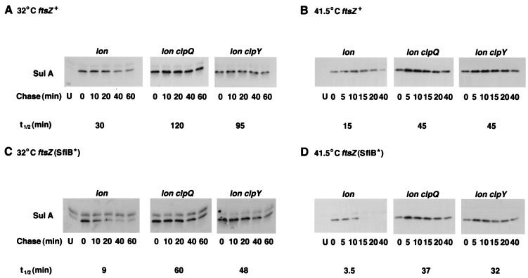
FIG. 3.
Turnover of SulA in lon clpQY mutants. Strains were grown at 32°C in LB and exposed to UV light as described in Materials and Methods. After dilution to the original volume in LB, growth was continued in the dark for 25 min at 32°C (A and C) or for 10 min at 41.5°C (B and D). Spectinomycin (150 μg/ml) was added to inhibit further protein synthesis, and samples were removed and processed for Western blotting with anti-SulA antibody as described in Materials and Methods. (A) ftsZ+ strains SG22186, SG22207, and SG22208 (panels from left to right) at 32°C. (B) ftsZ+ strains at 41.5°C. (C) ftsZ(SfiB*) strains SG22224, SG22225, and SG22226 (panels from left to right) at 32°C. (D) ftsZ(SfiB*) strains at 41.5°C. Lanes U contained lon mutant hosts sampled before UV induction (uninduced) to indicate the position of the UV-inducible SulA band. Half-lives (t1/2) were determined by scanning of the Western blots. Because many of the 5-min spots showed more protein than 0-min samples, presumably due to the delayed action of spectinomycin, the half-lives were calculated with the 5-min samples as time-zero samples.

