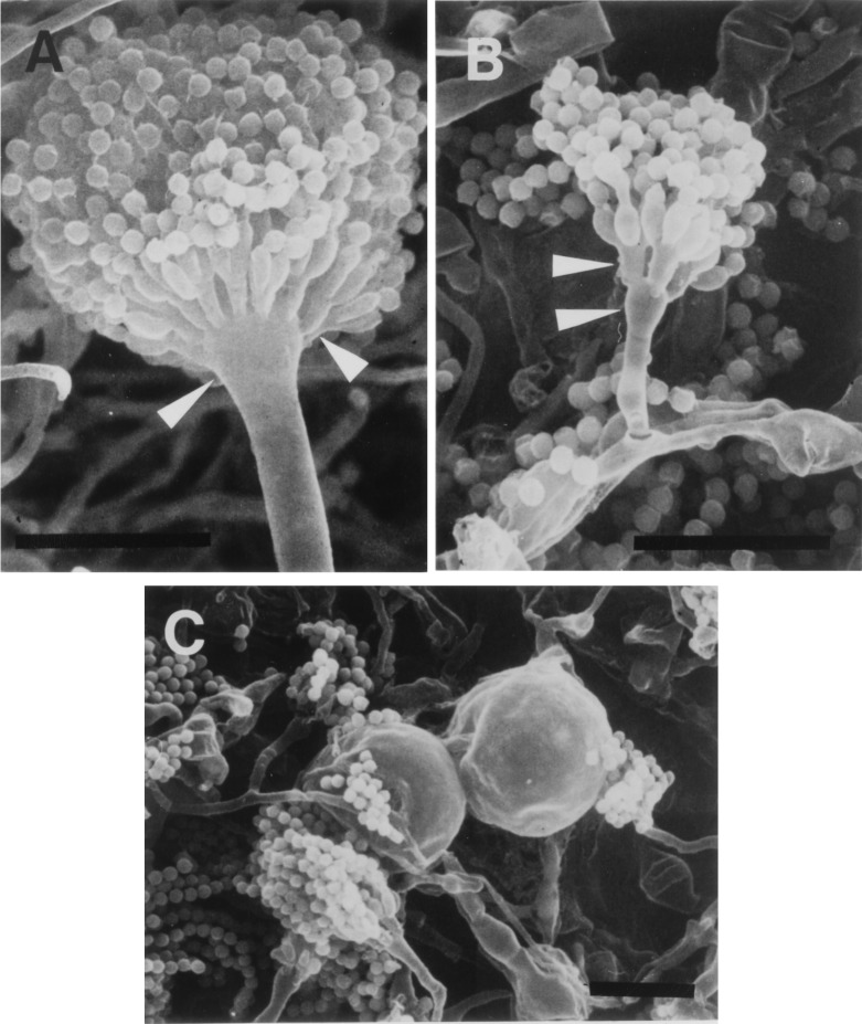
FIG. 5.
Scanning electron microscopic images of mycelia and conidiophores of the ΔcsmA mutant. Panels: A, ABPU/A1; B and C, M-9. Conidiophore structure abnormality was found in the short stalk, small vesicle (lower arrowheads), and a small population of metulae (upper arrowheads). Mycelia cultured for 7 days were used. Bars, 20 μm.

