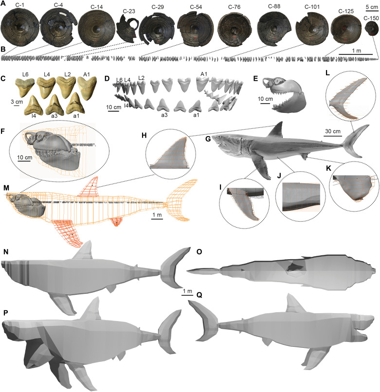
Fig. 1. Modeling procedure.
(A) Sample of 11 of the 141 vertebral centra in the Otodus megalodon column (IRSNB P 9893). (B) 3D scan and reconstruction of the O. megalodon vertebral column, with centra from (A) linked to their corresponding position. (C) Sample of seven O. megalodon teeth from the UF 311000 dentition (lingual view) with their respective positions (uppercase denotes upper teeth; lowercase refers to lower teeth; “A” denotes anterior teeth, and “L” lateral). (D) 3D scan and reconstruction of the UF 311000 dentition (labial view) with the corresponding labels from (C). (E) 3D scan of Carcharodon carcharias chondrocranium used to model O. megalodon’s head. (F) C. carcharias chondrocranium with UF 311000 dentition and IRSNB P 9893 column attached and hoops outlining the model’s head. (G) 3D scan of the full body of the C. carcharias specimen used for flesh reconstruction with elliptical hooping methodology indicated for the (H) dorsal fin, (I) pectoral fins, (J) abdomen, (K) pelvic fins, and (L) caudal fin. (M) Base skeletal model with octagonal hoops that mark flesh boundaries. (N) Final lofted polygon mesh of O. megalodon used for analyses at lateral view and (O) dorsal view. (P) Visualization of open gape at 75° angle at oblique view and (Q) 35° gape angle at lateral view.

