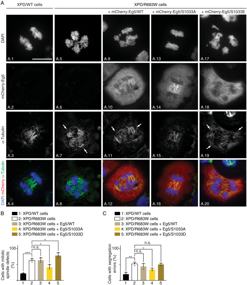
Fig. 7. Eg5/S1033A restores mitotic defects in XPD-mutated patient cells.
(A) Immunofluorescence of XPD/WT and XPD/R683W cells overexpressing either tagged mCherry-Eg5/WT, mCherry-Eg5/S1033A, or mCherry-Eg5/S1033E in anaphase. Cells were synchronized in mitosis for 16 hours with nocodazole (100 ng/ml) and collected 90 min after nocodazole release. Immunofluorescence analyses were performed with antibodies targeting the mCherry Tag and the mitotic spindle marker α-tubulin. Chromosomes were stained with DAPI. Arrows point to DNA bridges. Scale bar, 5 μm. (B and C) Percentage of cells displaying mitotic spindle defects (B) and segregation errors (C) (n = 3, means ± SD; at least 300 cells per experiment and per condition were counted; *P < 0.05, **P < 0.01, and ***P < 0.001, Student’s t test).

