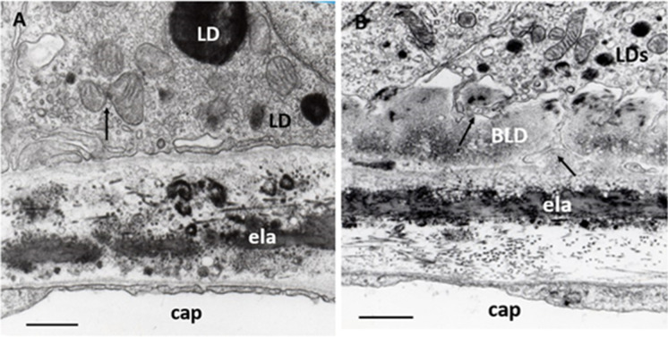
Fig. 5.
BLD in early AMD. A Normal aged Bruch’s membrane. RPE contains some small mitochondria, two of them show fission (arrow) and electrodense LDs; inner and outer collagenous layers contain electrodense inclusions of various shapes and sizes, as well as some of them are round and single membrane-bound; elastic layer, basal lamina of RPE, and capillary endothelium are normal. ela: elastic layer, cap: choriocapillaris. Seventy-four years; male; ×28,000; bar: 1 μm. B BLD in early AMD. A longitudinal section of BLD between the basal lamina and basal cytoplasm of RPE. In some places, it is amorphous in appearance with small electron-translucent vacuoles, while in other palaces it shows filamentary structures with electron-dense patches due to lipids. Basal in-foldings of the RPE in some places may form deep protrusions into the BLD, reaching the basal lamina of the RPE (black arrows). The cytoplasm of RPE contains numerous small LDs and only a few mitochondria of various sizes but well-preserved cristae and matrix. ela: elastic layer, cap: choriocapillaris. Seventy-one years; female; ×25,000; bar:1 μm

