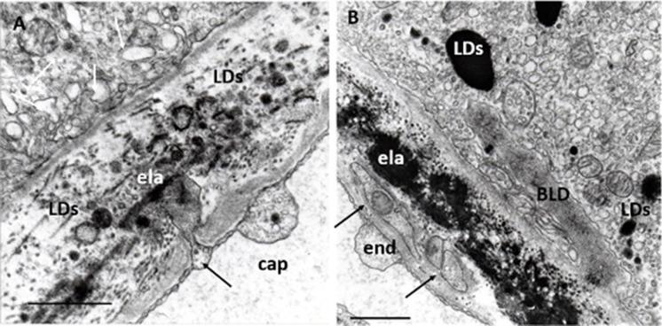
Fig. 8.
Endothelial alterations. A Endothelial sprouting in a normal eye. Small, deep sprouting reaches the elastic layer of the Bruch’s membrane (black arrow), which contains several LDs of various sizes and shapes can be seen. Numerous dilated lysosomes and small round-form lysosomes next to a mitochondrion can be seen in the RPE (white arrows) ela: elastic layer; 48 years; female; ×32,000; bar: 1 μm. B Endothelial cell processes of in early AMD. Transversal section of endothelial cell processes in the basal lamina of the choriocapillaris (arrows). They contain small mitochondrion with confluent cristae. BLD, some small sparsely distributed mitochondria, several lysosomes, and LDs can be seen in the RPE. ela: elastic layer. Eighty-four years; female; ×22,000; bar: 1 μm

