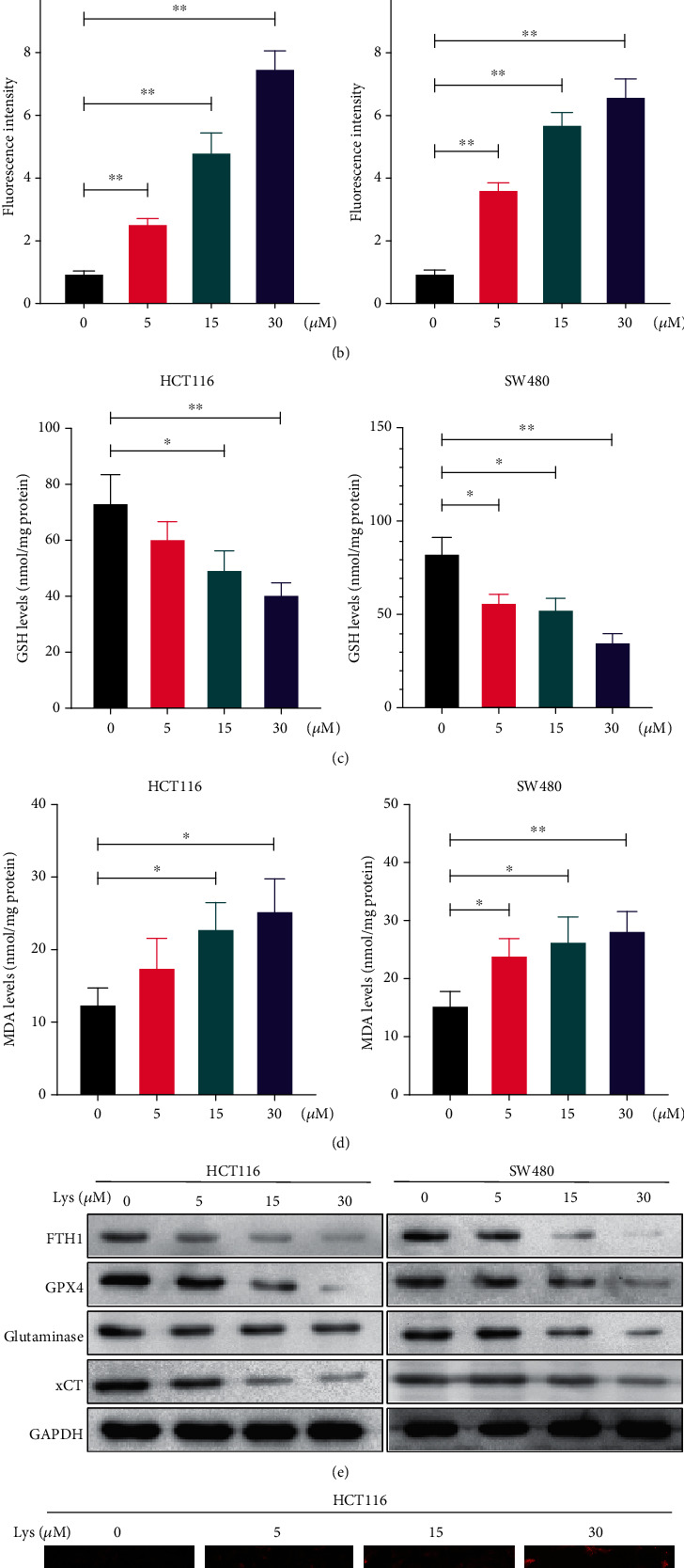
Figure 3.

Lys triggered ferroptosis in CRC cells. (a, b) ROS levels were measured by DCFH-DA staining after HCT116 and SW480 cells had been treated with Lys for 24 hours. The results are shown as the mean standard deviation. (c) The GSH level in HCT116 and SW480 cells was determined after the treatment with Lys for 24 hours, and the results showed that there was a significant difference between the groups. (d) The MDA level in HCT116 and SW480 cells was measured following the treatment with Lys for 24 hours. (e) Following a treatment with Lys for 24 hours, the iron content of HCT116 and SW480 cells was evaluated using the ferroOrange staining method. (f) Western blotting was used in order to identify a number of proteins connected to ferroptosis. ∗P < 0.05 and ∗∗P < 0.01.
