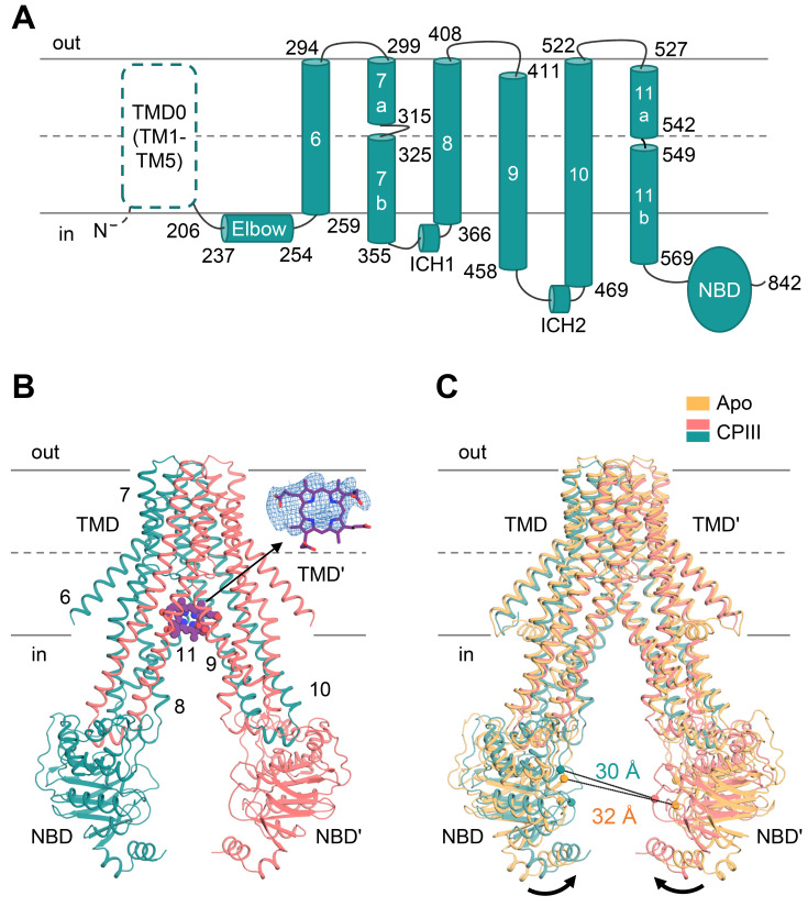
Fig. 2. Structure of CPIII-bound ABCB6-∆TMD0.
(A) Schematic representation of the human ABCB6 monomer. The N-terminal TMD0 was not included in the construct, and is drawn with a dashed line. (B) The molecular structure of CPIII-bound ABCB6-∆TMD0. The monomers are shown in pink and teal, respectively. The CPIII is shown as purple spheres. The density corresponding to CPIII (front view) is shown at 4 σ level. The elbow helix was not built due to poor electron density. Single apostrophes are used for the residues (or helices) of one monomer to differentiate them from those of the other monomer. (C) Structure comparison of apo (PDB ID 7D7R) and CPIII-bound ABCB6-∆TMD0. The Cα distance between the conserved G626 of the Walker A motif and the S728 of the signature motif is indicated. Black arrows indicate movements of the NBDs.

