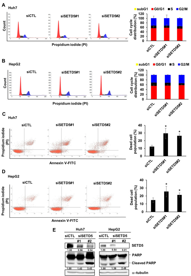
Fig. 3. SETD5 KD induces Huh7 and HepG2 cell apoptosis.
(A and B) Left: Effects of SETD5 KD in Huh7 and HepG2 cells on the cell cycle were examined by FACS analysis to detect propidium iodide signals. Right: Quantification of cell cycle distribution as determined using a flow cytometer. Values are the mean ± SEM of biological triplicate experiments. (C and D) Left: Effects of SETD5 KD in Huh7 and HepG2 cells on cell death were examined by FACS analysis based on annexin V and propidium iodide double staining. Right: Quantification of the dead cell population as determined using a flow cytometer. Values are the mean ± SEM of biological triplicate experiments (n = 3, *P < 0.05, one-way ANOVA). (E) Cleaved PARP in Huh7 and HepG2 cells was examined by immunoblotting with anti-PARP antibody. The intensities of immunoblotting signals were semiquantitatively measured by ImageJ.

