Fig. 2.
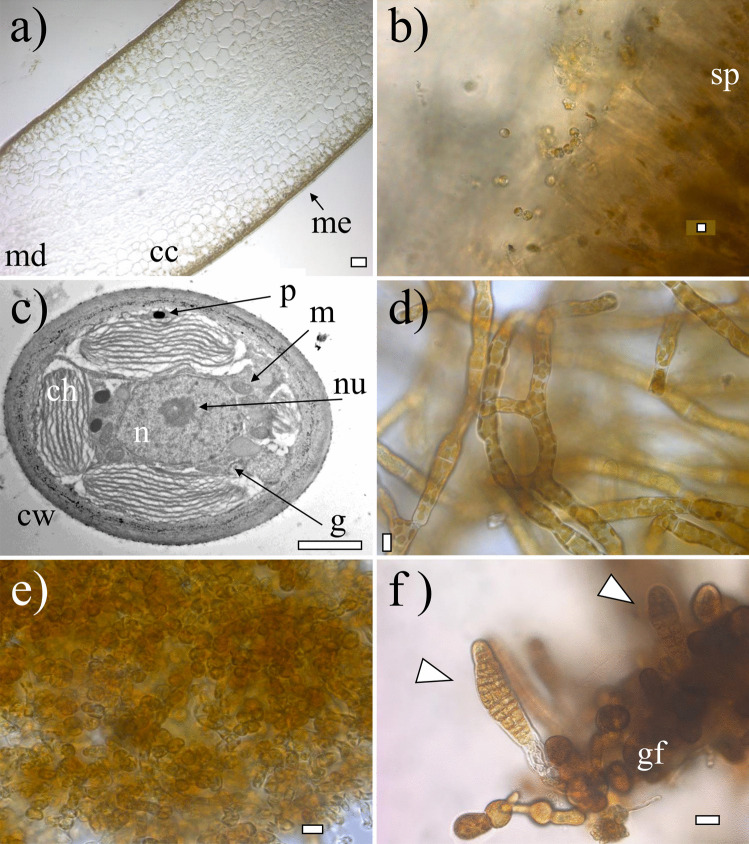
Morphology and anatomy of Saccharina latissima. a Transversal cross-section of an adult sporophyte showing the meristoderm (me), cortical cells (cc), and medulla (md); b fresh released zoospores from sporangia (sp) from a mature sporophyte; c transmission electron microphotograph of a gametophyte cell showing nucleus (n), nucleolus (nu), chloroplasts (ch), cell wall (cw), golgi bodi (g), mitochondria (m), and physodes (p); d female gametophyte culture; e male gametophyte culture; f early-stage embryonic sporophytes (indicated with triangles) still attached to female gametophyte (gf). Scale bar: a 50 µm, b 5 µm, c 2 µm, d–e 10 µm, and f 20 µm

