Fig. 3.
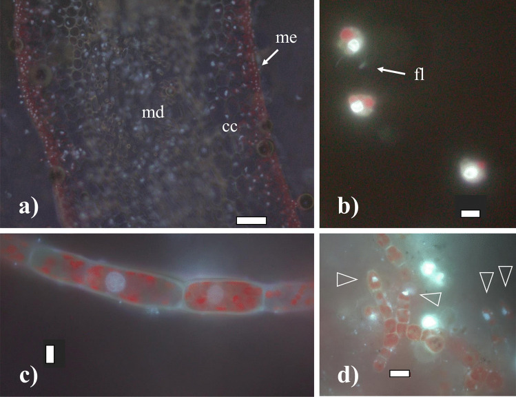
Cells of Saccharina latissima under fluorescence, stained with DAPI without methacarn treatment showing chloroplasts in dark red and stained nuclei in light white/blue: a transversal cross-section of an adult sporophyte showing the meristoderm (arrow: me), cortical cells (cc) and medulla (md); b released zoospores with chloroplasts, a nuclei and flagella (arrow: fl); c female gametophyte filament with cylindrical cells chloroplasts; d male gametophyte with rounded cells and many chloroplasts, nuclei indicated with triangles. Scale bar: a 100 µm, b–c 5 µm, and d 10 µm

