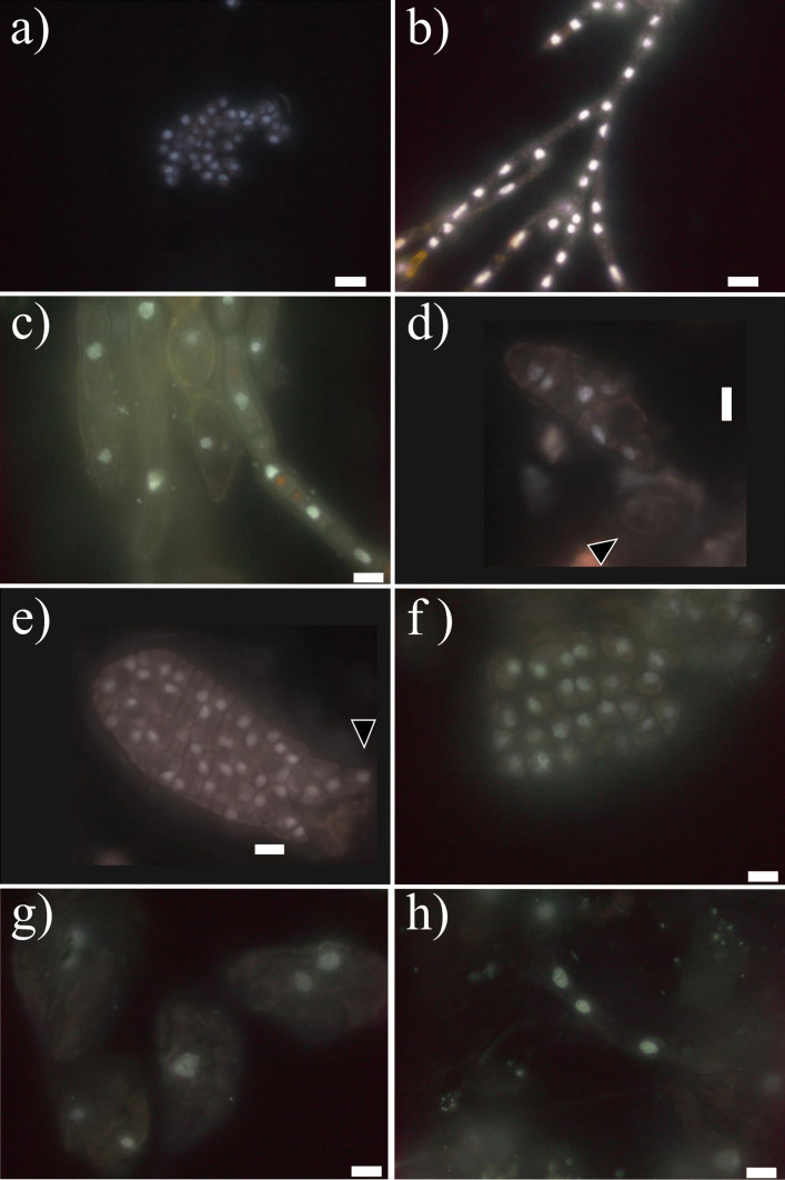
Fig. 5.
Different DAPI-stained nuclei after methacarn treatment: a from released zoospores; b from male gametophytes; c different forms and sizes from female gametophytes; d–e from early-stage development of embryonic sporophytes including a few celled sporophyte still attached to the female gametophyte (black arrow, d) and a further developed sporophyte with rhizoidal cells (black triangle, e); and f–h from different tissue cells of full developed sporophyte including meristoderm (f) and large cortical (g) and long medullar cells (h). The scale bar in all pictures corresponds to10 µm

