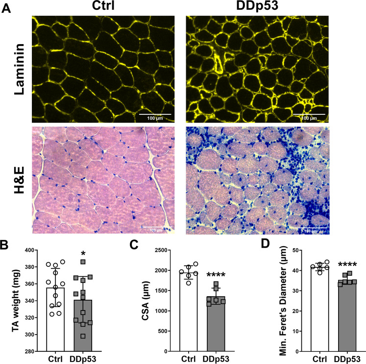
Fig. 2. Changes to muscle mass, fiber size and health after DDp53 electroporation.
A Immunohistological assessment of myofiber cross-sectional area via laminin staining (Laminin, upper row), and hematoxylin and eosin staining (H&E, lower row) to assess fiber integrity indicated central nuclei, regenerating fibers, macrophage infiltration, and fiber death. B Muscle mass in the DDp53 tibialis anterior was significantly reduced three weeks after electroporation. C Fiber cross sectional area was significantly reduced in DDp53 three weeks after electroporation. D Fiber diameter was significantly reduced in DDp53 three weeks after electroporation. n = 6 to 12, * indicates a p-value of <0.05, **** indicates a p-value of <0.0001.

