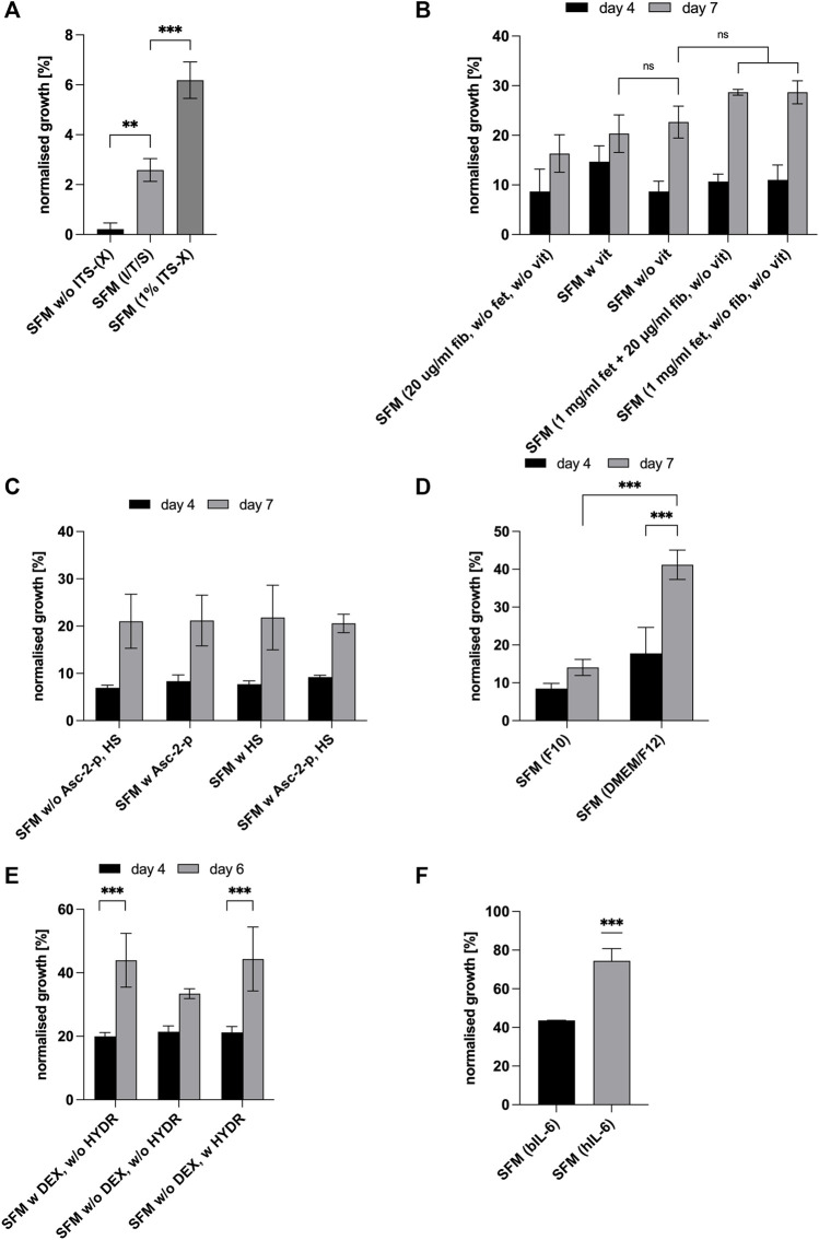
FIGURE 2.
For all graphs, cell growth was measured with MTS or HCA and expressed as percentages of GM (control, not shown in graph). (A) Effect of the addition of insulin (I), transferrin (T) and sodium selenite (S) in comparison to supplementation of SFM with commercial Gibco® ITS-X (X = Ethanolamine) at day 7 of culture. Asterisks (**p < 0.01, ***p < 0.001) indicate significant effect of the presence of ITS (-X) in the serum-free formulation [SFM w/o ITS (-X)]. (B) Effect of attachment factors [vitronectin (v), fibronectin (fib) and fetuin (fet)] at different concentrations on cell growth in SFM and expressed as % of growth achieved in GM (bars not shown) on days 4 and 7. (C) Effect of L-Ascorbate-2-phosphate and heparan sulphate on cell proliferation on days 4 and 7. For all conditions, time significantly impacted cell proliferation (p < 0.001). (D) Effect of different basal media and time, DMEM/F-12 and Ham’s F-10 Nutrient Mix on cell expansion on days 4 and 7. Asterisks (***p < 0.001) indicate that the mean value is significantly different between DMEM/F12 & F10 and that there is a significant time effect for DMEM/F12. (E) Effect of dexamethasone and hydrocortisone on cell proliferation on days 4 and 6. Asterisks (***p < 0.001) indicate that there is a significant time effect for—DEX + HYDR. (F) Comparison of bIL-6 with hIL-6 on days 4 and 6. SFM with hydrocortisone and DMEM/F12 in both cases.

