FIGURE 8.
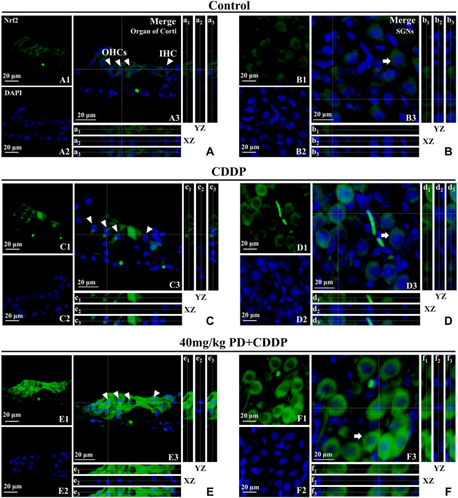
PD promotes Nrf2 translocation into the nucleus and increases HO-1 expression. Arrowheads indicate OHCs and IHC; arrows indicate SGNs. (A–F): representative immunofluorescence labelling in the organ of corti (left panel) and SGNs (right panel) for Nrf2 (green), double stained with DAPI in blue. A3-F3 represent the merged images. X–Z and Y–Z cross-sections in boxes (referred to the dashed lines) from the Z-stack acquisitions show cytosolic or nuclear fluorescence signal (XZ and YZ boxes, a1-f1: Nrf2 fluorescence; a2-f2: DAPI staining; a3-f3: Merge). Cisplatin administration (C,D) induced an antioxidant response in the cochlea, resulting in a slight increase of Nrf2 expression in the cytoplasm [XZ and YZ c1-c3 and d1-d3 in (C,D)] compared to the control group [XZ and YZ A1-A3 and B1-B3 in (A,B)]. After PD administration (E,F), the expression of Nrf2 was further enhanced in the cytoplasm and Nrf2 translocated to the nucleus [XZ and YZ e1-e3 and f1-f3 in (E,F)]. Scale bar, (A–F), 20 µm.

