Figure 4.
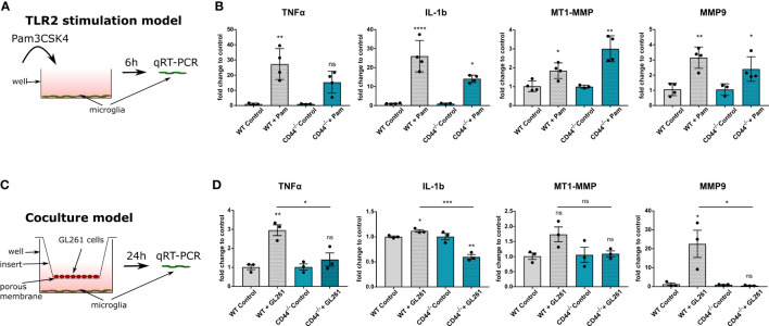
CD44 on myeloid cells is implicated in TLR2 signaling and MMP9 expression. (A) Schematic representation of the experimental setup. Primary microglia isolated from control or CD44-/- mice were stimulated with 10ng/ml of the TLR2 agonist Pam3CSK4. After 6 hours, total RNA was extracted from the cells and processed for qRT-PCR analysis. (B) mRNA levels of TNF-α, IL-1b, MT1-MMP and MMP9 after 6 hours of Pam incubation. Bar charts represent mRNA levels as fold change to the corresponding control condition. Data are presented as mean ± SD. Student’s t-Test, ****p < 0.0001, **p < 0.01, *p < 0.05 vs corresponding control. (C) Schematic representation of the co-culture experimental setup. Primary microglia isolated from control or CD44-/- mice were cultured in 6-well plates. GL261 cells were seeded in the top compartment on 0.4 µm porous inserts. For control condition, no cells were seeded on the insert. Co-culture was then carried-out for 24 hours after which total RNA was extracted from microglia and processed for qRT-PCR analysis. (D) mRNA levels of TNF-α, IL-1b, MT1-MMP and MMP9. Bar charts represent mRNA levels as fold change to the corresponding control condition. Data are presented as mean ± SD. Student’s t-Test, **p < 0.01, *p < 0.05 vs corresponding control. n.s., not-significant.

