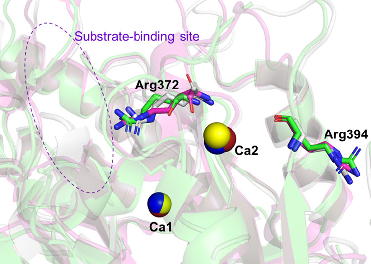
Figure 10.

Structures around Arg372, which is citrullinated in PAD2, PAD3, and PAD4, and Arg394, which is citrullinated in PAD3 and PAD4.28,30 The structures of PAD2, PAD3, and PAD4 are all Ca2+-bound forms and colored in gray, green, and magenta, respectively. Only Arg372 and Arg394 (PAD3 numbering) are depicted in stick models, in which the N and O atoms are colored in blue and red, respectively. The Ca2+ (Ca1 and Ca2) ions bound in the vicinity of the two arginine residues of interest are depicted as red, yellow, and blue spheres in the PAD2, PAD3, and PAD4 structures, respectively. The purple dotted circle indicates the vicinity of the substrate-binding site. PAD, peptidylarginine deiminase.
