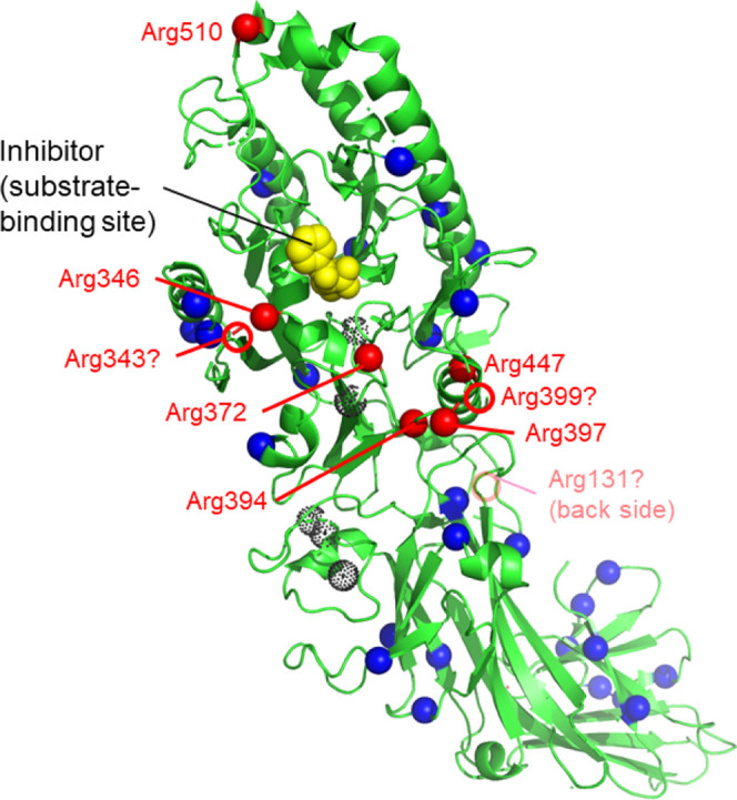
Figure 7.

Distribution of arginine and citrullinated arginine residues in PAD3. The structure of WT PAD3-Ca2+-Cl-amidine (Chain A of PDB ID; 7D56) is shown in green. The dotted black spheres indicate Ca2+ ions. Blue and red spheres indicate arginine and autocitrullinated arginine residues, respectively. In 7D56, six of the nine citrullinated arginine residues were visualized. Red circles indicate three autocitrullinated arginine residues (Arg131, Arg343, and Arg399) at putative positions that were not identified following the X-ray crystallographic structure analysis. The inhibitor Cl-amidine is shown in yellow. Peptidylarginine deiminase 3 (PAPD3); wild type (WT).
