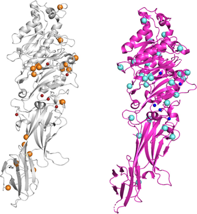
Figure 9.

Potential autocitrullinated sites of PAD isozymes. (Left) Structure of PAD2-Ca2+ (PDB ID; 4N2C), depicted in a gray cartoon model. The small red and large orange spheres represent Ca2+ ions and arginine residues which are supposed to be autocitrullinated, respectively. (Right) Structure of PAD4-Ca2+ (PDB ID; 1WD9), depicted in a magenta cartoon model. The small blue and large cyan spheres represent Ca2+ ions and the arginine residues which are supposed to be autocitrullinated, respectively. Unlike the distribution of citrullinated arginine residues in PAD3 (shown in Figure 7), the arginine residues to be citrullinated appear to be uniformly distributed throughout the structure of PAD2-Ca2+ and PAD4-Ca2+. Peptidylarginine deiminase (PAD); wild type (WT).
